Loading…
Chest radiograph, PA view
Plain radiograph · Posteroanterior, erect · Thorax
Before you comment on anything
This is the film every doctor reads more often than any other, and the one students rush. Nobody is asking you to spot a subtle nodule in your first year. They are asking whether you read a film in a fixed order instead of staring at the middle. Say the modality and the view first, prove the film is adequate, then work outwards from the airway. If you do that on a normal film you will still do it on an abnormal one.
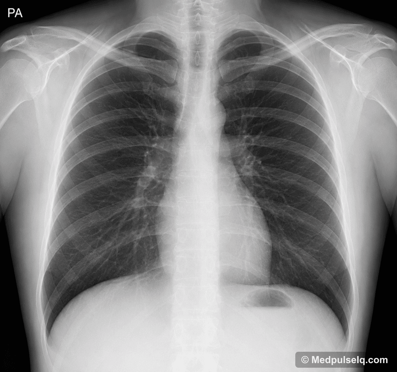
Plain radiograph · Posteroanterior, erect · © MedpulseIq.com
Read it in order
0 of 8
Work down the list. The answer unlocks when you have been through all of it.
Landmarks to name
Trachea and carina
In the midline of the upper mediastinum, dividing at about the level of the fifth thoracic vertebra
Deviation is the quickest sign that something is pushing or pulling the mediastinum.
Right and left hilanot marked on this film
Either side of the mediastinum at mid-chest, formed mainly by the pulmonary arteries
The left hilum normally lies slightly higher than the right; the reverse is always abnormal.
Cardiac silhouette
Mostly to the left of the midline
The right border is the right atrium and the left border the left ventricle, so the silhouette is an anatomical map.
Costophrenic anglesnot marked on this film
Where each hemidiaphragm meets the chest wall laterally
The most dependent part of the erect pleural cavity, so the first place fluid collects.
Right and left hemidiaphragmsnot marked on this film
Curving domes at the lung bases
The right sits higher because of the liver; gas below the right dome is never normal.
Viva voce
1 of 5 · 0 answeredSay your answer out loud before you open it. That is the exam skill.
Why is a chest radiograph taken posteroanteriorly rather than anteroposteriorly?
Senior’s 30-second takeaway
Chest radiograph, PA view — the whole bench in four lines. Read this on the way in.
- If you see this
- A chest film with the scapulae clear of the lung fields · A central trachea and sharp costophrenic angles · A heart occupying less than half the chest width
- If asked to identify it
- This is an adequate posteroanterior erect chest radiograph and it is normal.
- If asked one more
- Adequacy is rotation, inspiration and projection, and it is stated before any comment on the lungs.
- If asked why it matters
- Free gas under the right hemidiaphragm on an erect film means a perforated viscus until proved otherwise.
Then stop. Let the examiner ask the next question.