Loading…
Gallbladder
Spotter answer: T.S. of gallbladder
Clear field
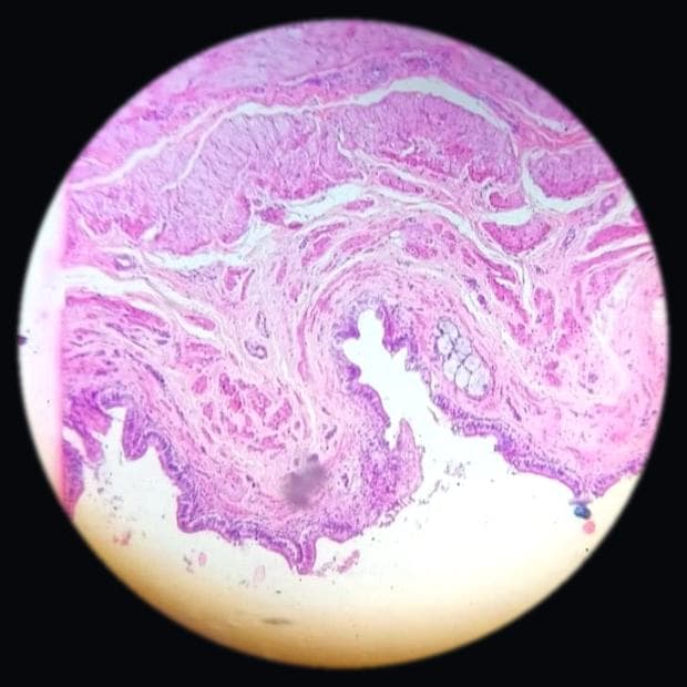
Highly folded mucosa of gallbladder at medium power. Adjust the focus1.0×
- 1Folded mucosa — Increases surface area for bile concentration.
- 2Simple columnar epithelium — Lines the mucosal folds.
- 3Lamina propria — Supports the epithelium.
Before you name anything
Take a deep breath and let's start by looking at the overall structure of the slide. At low power, you'll notice the gallbladder's mucosa is highly folded, which is a key feature. This folding increases the surface area and is lined by simple columnar epithelium. As you move in closer, you'll see there's no muscularis mucosae or submucosa, which is distinctive. The wall is relatively simple, with a lamina propria and an irregular smooth muscle layer. Many students make the mistake of confusing this with the small intestine due to the folds, but remember, the gallbladder lacks the villi and crypts found in the intestine. Focus on the simplicity of the wall and the absence of certain layers to guide you. This is your way in: identify the folds, confirm the epithelium, and note the absence of muscularis mucosae and submucosa. Keep calm, and you'll do just fine.
Sign in free to open the rest of this slide
The identification ladder, the marks-earning features, the look-alikes, the record-book diagram and the five viva questions are waiting. Free to start, no payment.
Sign in free