Loading…
Histology practical
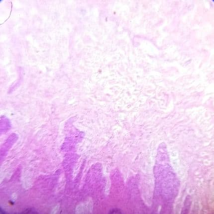
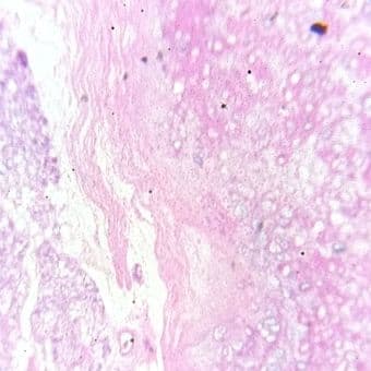
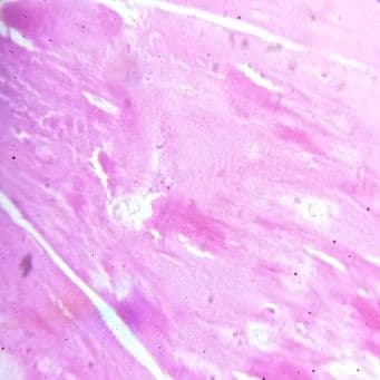
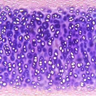
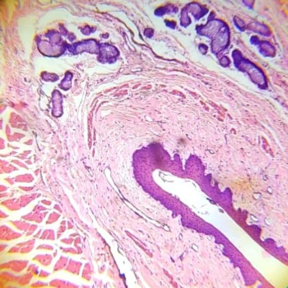
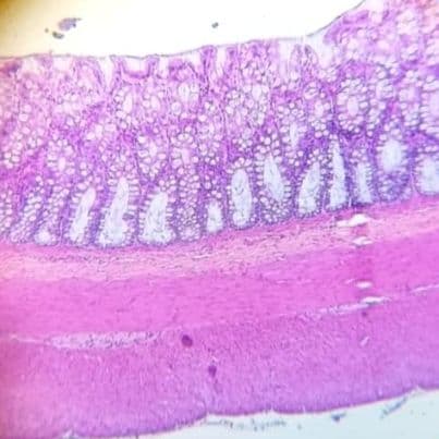
50 specimens across 11 organ systems, photographed down a student microscope — the same slides, the same lighting, the same imperfect fields you will meet in the lab. Each one comes with a senior talking you through it: what to do at low power, the two or three features that earn marks, the look-alike that catches people out, and the viva that follows.
- Specimens
- 50
- Photographed fields
- 68
- Viva questions
- 250
- Spotter slides
- 64
The first 10 specimens are open to everyone. Sign in free to open the rest and keep your progress.
Your practical readiness
0 of 50 specimens you can nameNothing marked yet — open any slide and tick it once you can name it without help. Kept on this device only.
- General histology0/11
- Oral cavity & GIT0/11
- Glands & hepatobiliary0/5
- Lymphoid organs0/4
- Urinary system0/3
- Male reproductive0/3
- Female reproductive0/3
- Endocrine system0/3
- Respiratory system0/2
- Nervous system0/3
- Special senses0/2
Compare bench
The look-alikes. Find the difference before you turn the names on.
Three tubes with villi — the submucosa settles it.
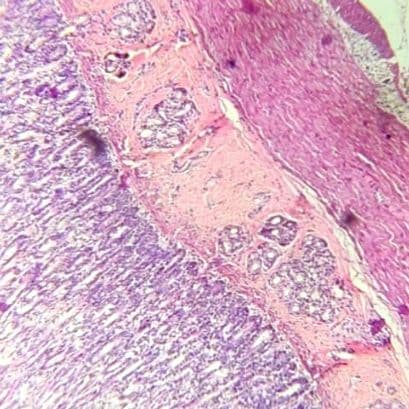
Specimen 1
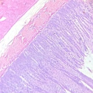
Specimen 2
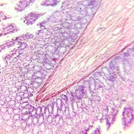
Specimen 3
General histology
11 specimens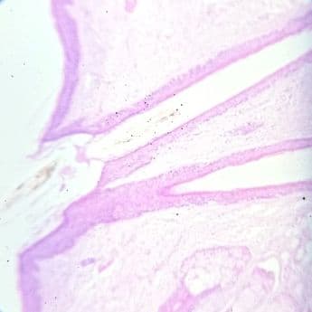
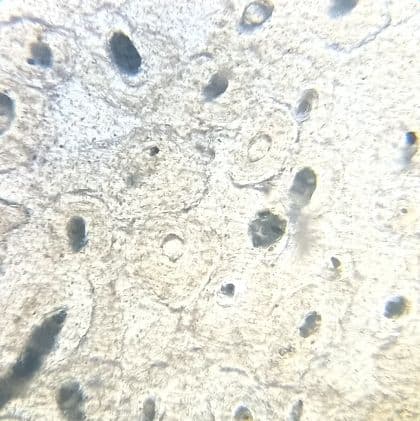

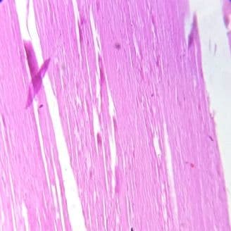
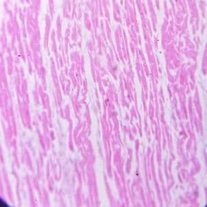
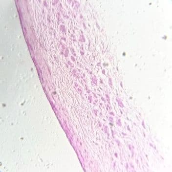
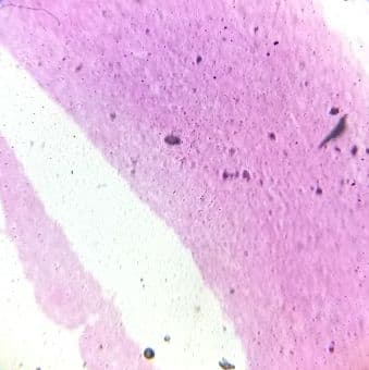
Oral cavity & GIT
11 specimens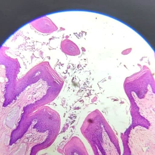
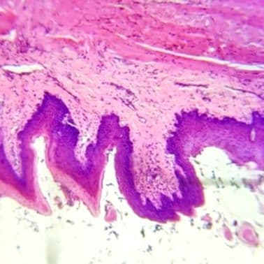
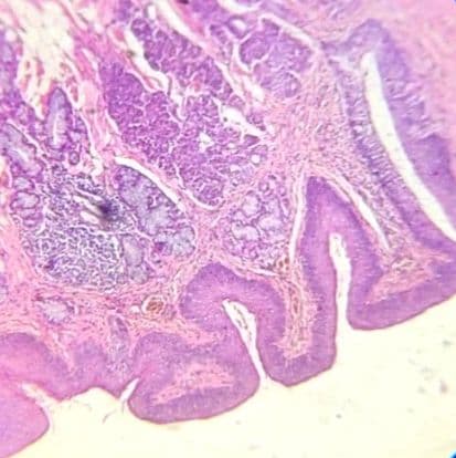
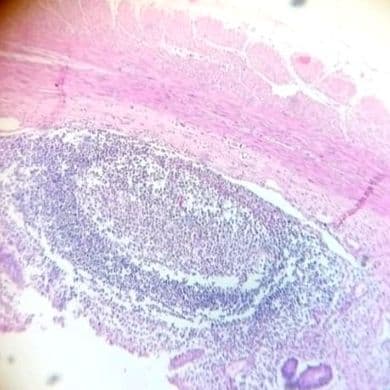
Glands & hepatobiliary
5 specimens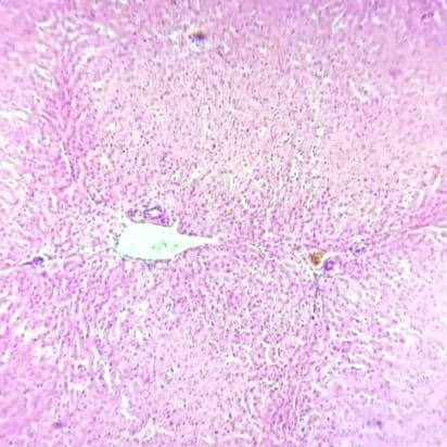
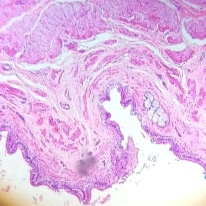
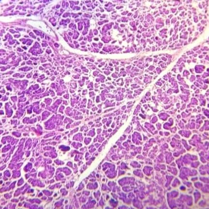
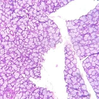
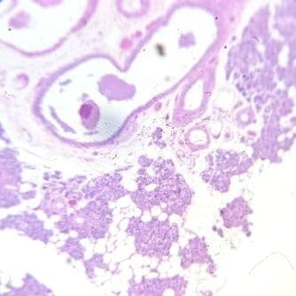
Lymphoid organs
4 specimens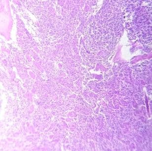
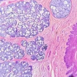
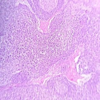
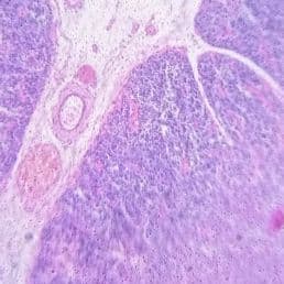
Urinary system
3 specimens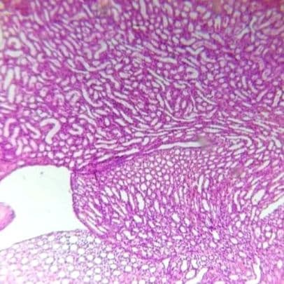
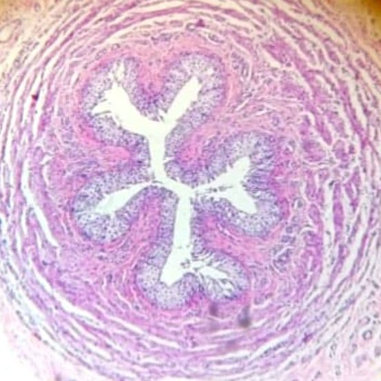
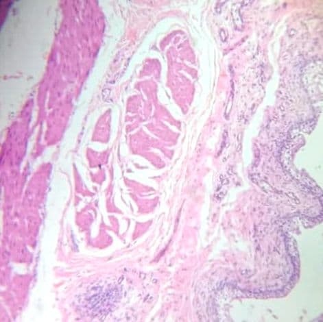
Male reproductive
3 specimens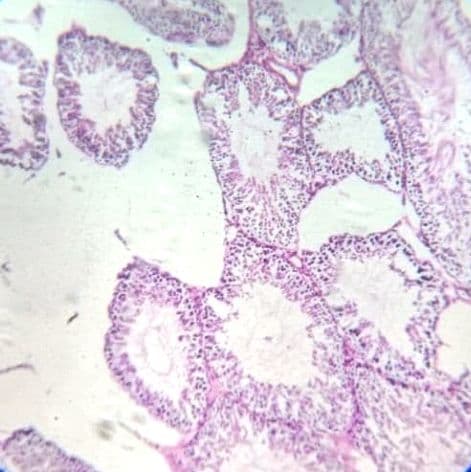
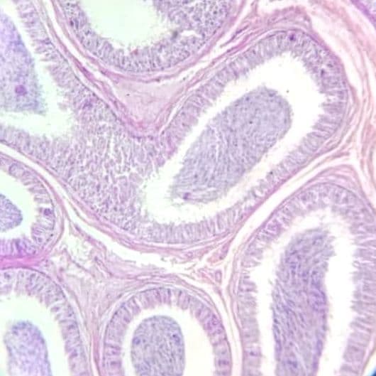
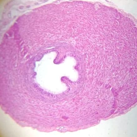
Female reproductive
3 specimens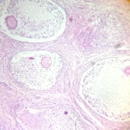
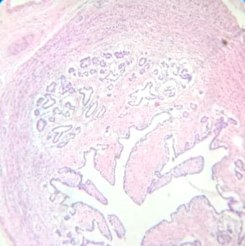
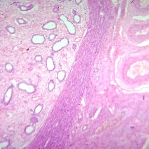
Endocrine system
3 specimens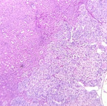
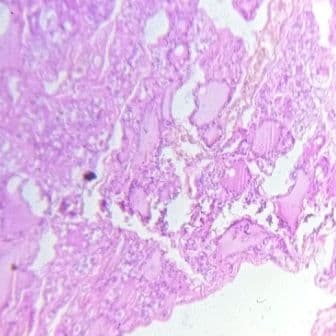
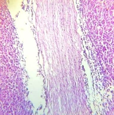
Respiratory system
2 specimens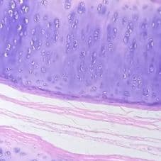
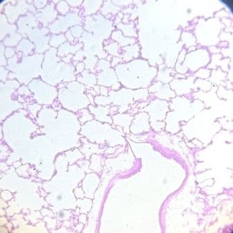
Nervous system
3 specimens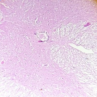
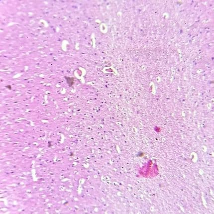
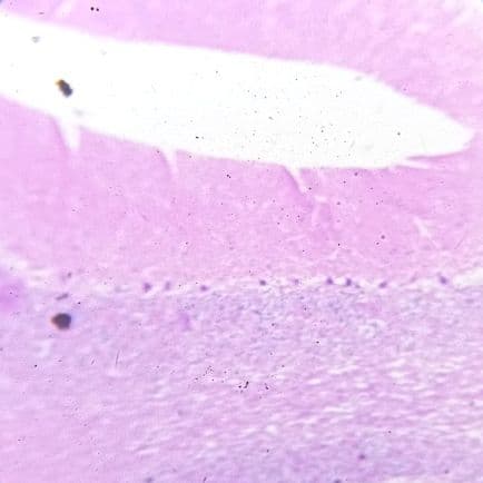
Special senses
2 specimens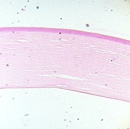
About these photographs
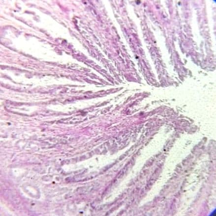
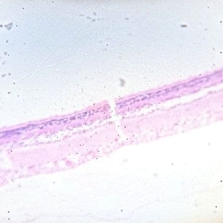
These are real fields photographed down a teaching microscope and contributed for MedPulse IQ, not textbook plates or whole-slide scans. That is deliberate — it is what your slide will actually look like. Where a field is too washed out to identify from, we say so on the slide and keep it out of every quiz rather than pretend.
1 more specimens are still with review. Every specimen here has been checked against its own photograph by a second reader; anything that did not pass is held back rather than published.
Fields we want to re-shoot (4): Bone — longitudinal section, Large vein, Large vein, Large artery.