Loading…
Hyaline cartilage
Spotter answer: Hyaline cartilage section
Clear field
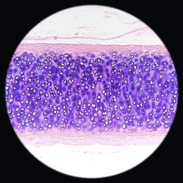
Hyaline cartilage at medium power showing visible chondrocyte nuclei. Adjust the focus1.0×
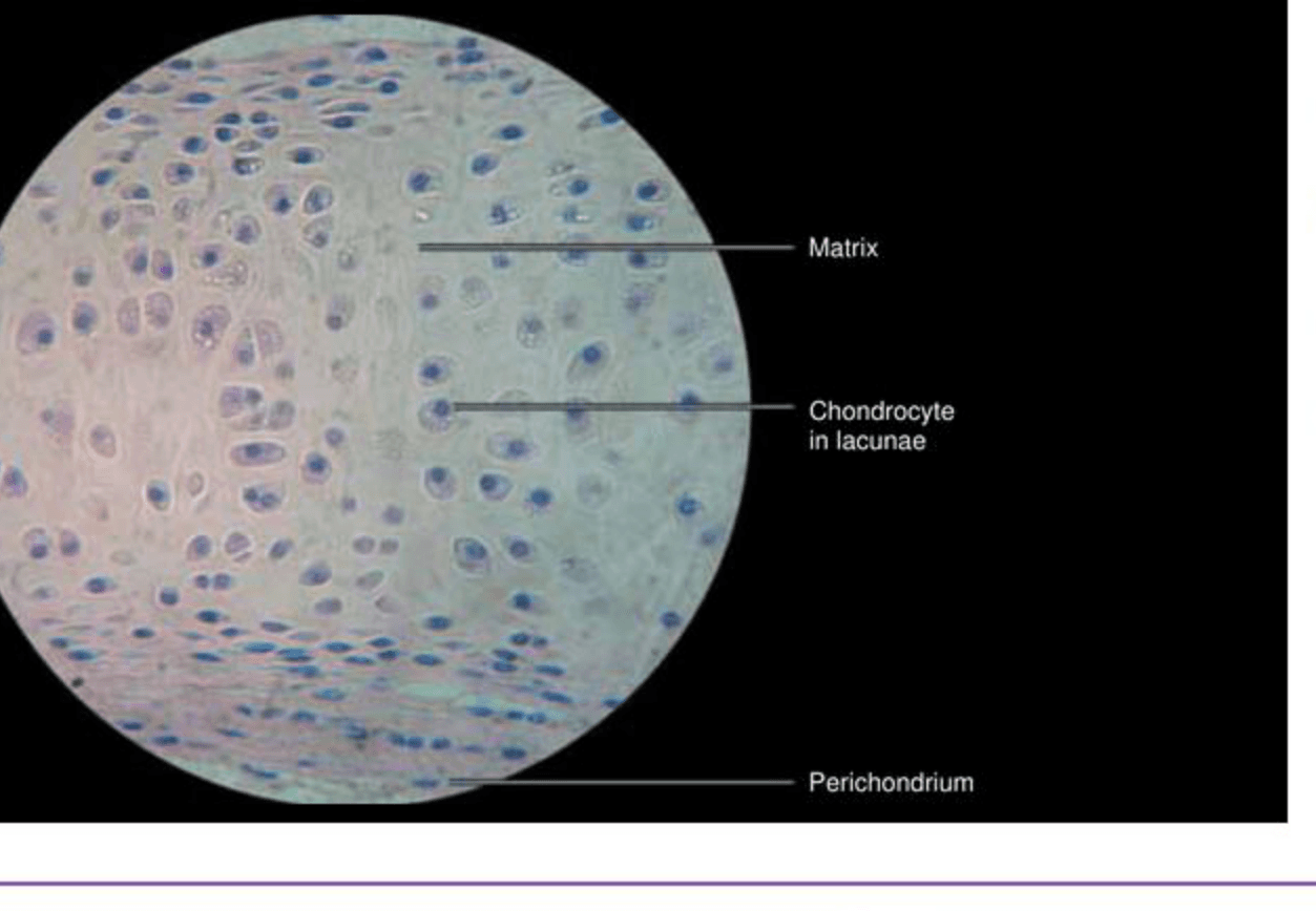
- 1Chondrocytes — These are the cells within lacunae.
- 2Lacuna — Space housing the chondrocyte.
- 3Perichondrium — Dense connective tissue covering the cartilage.
Hyaline cartilageIllustration, not a specimen
Before you name anything
Take a deep breath and let's start by looking at the whole field at low power. Notice the overall shape and texture. You’re seeing a smooth, glassy matrix, typical of hyaline cartilage. Focus on the lacunae, which house the chondrocytes. These are often in small groups, known as isogenous groups. The matrix is relatively uniform, which is a key feature here. Students often rush to high power and miss the overall architecture, so stay calm and start broad. The presence of a perichondrium at the edge can help confirm your identification. Remember, the smooth matrix and the lacunae are your main clues.
Work it out, one rung at a time
Answer each question in your head first. Then open it.
- 1
low-power architecture
What is the overall shape and texture?
- 2
epithelium or covering
Is there a covering present?
- 3
the signature feature
What is the key feature of this tissue?
- 4
confirm and exclude the look-alike
How do you know it's not fibrocartilage?
Viva voce
1 of 5 · 0 answeredSay your answer out loud before you open it. That is the exam skill.
What cells are found in hyaline cartilage?