Loading…
🩻 Radiology · Part 10 revision hub
Integrated Interpretation and Practice
Everything you need to revise this Part in one place — the one‑page note for every concept, a rapid‑revision sheet, exam‑style questions that point you back to the right concept, flashcards, staged cases and viva practice.
5 concepts20 MCQs20 flashcards3 cases12 viva questions
📝 The plate wall
Every concept in this Part as a one‑page handwritten note. Open one to read it in full and zoom.
⚡ Rapid revision
The whole Part on one screen — read it the night before.
Interpretation
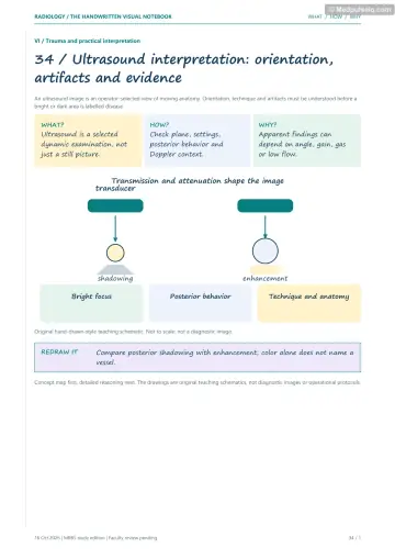
- US: reflection drives image formation. Anechoic = clear fluid (+ posterior enhancement); internal echoes = complex fluid (pus, blood); solid = add Doppler; hyperechoic + shadow = calculus/bone; hyperechoic + comet tail/ring‑down = gas.
- Discriminators: cystitis (echoes + thick wall), abscess (thick wall, echoes, fever), haemorrhagic cyst (lace), endometrioma (ground glass), dermoid (tip of the iceberg), calculus vs polyp vs sludge, bladder stone (mobile) vs tumour (fixed, vascular).
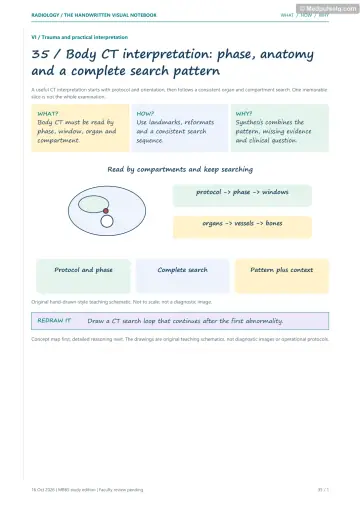
- CT: identify phase (bright aorta = contrast; excretory = collecting systems), windows, then history → region → organ → lesion → density → diagnosis.
- Density: air −1000, fat −50 to −100, simple fluid 0–20, exudate > 20, haemoperitoneum 30–45, clot 45–70 (sentinel clot), enhancement ≥ 20 HU rise, calcification > 100–200. Fat narrows the differential (AML, dermoid, lipoma, myelolipoma).
- CT pelvis limited: US first, MRI for detail.
Pathways, preparation and law
- US first: gallstones, effusion, HPS, intussusception, appendicitis (child), pregnancy, DVT, thyroid, breast < 30, FAST. CT: trauma, SAH, stroke first, CTPA, obstruction, NCCT KUB, pancreatitis at 72–96 h, CTA dissection, adult appendicitis, C‑spine. MRI: tumour, epilepsy, MS, cord, osteomyelitis, AVN, occult fracture, ligaments, MRCP, rectal/cervical staging, prostate. Nuclear: DMSA, MAG3, Meckel's, Tc‑RBC, bone scan, HIDA. Unstable patient: treat first.
- Preparation: GB US fasting; pelvic US full bladder; barium meal NBM 6 h; barium enema/CTC bowel prep; HSG days 7–10; mammography no deodorant/talc; FDG PET fasting + glucose; contrast CT: allergy, eGFR, metformin; MRI safety screening; intervention: consent + coagulation.
- Report: clinical question → technique → findings → impression (answer + differential) → recommendations (guideline systems). Critical findings phoned and documented.
- AERB: licensing (eLORA), RSO, TLD monitoring, dose limits, QA. PC‑PNDT: register machines, Form F, no sex disclosure. MTP Act 2021: 20 wk (one provider), 20–24 wk special categories, > 24 wk fetal anomaly (medical board). DPDP Act 2023: data confidentiality.
Named signs
- For every sign: what you see → mechanism → classic cause → differentials → next step (the 4S read).
🎯 Single‑best‑answer MCQs
Pick an answer to see the explanation — and the concept to revisit if you missed it. Your answers are saved on this device.
Score 0 / 00 of 20 answered
Question 1
The most important physical process in ultrasound image formation is:
Question 2
A hyperechoic focus with a "dirty" shadow and comet‑tail artefact most likely represents:
Question 3
On ultrasound, a bladder calculus is distinguished from a bladder tumour by:
Question 4
On a contrast‑enhanced CT, which feature indicates the image was acquired after IV contrast?
Question 5
A free intraperitoneal fluid collection measuring 55 HU adjacent to the spleen after trauma suggests:
Question 6
The investigation of choice for suspected common bile duct stones without ionising radiation is:
Question 7
A patient with suspected tension pneumothorax and hypotension should first undergo:
Question 8
The investigation of choice for suspected osteomyelitis in a child with a normal radiograph is:
Question 9
The investigation of choice for local staging of rectal cancer is:
Question 10
Patient preparation for a transabdominal pelvic ultrasound requires:
Question 11
Hysterosalpingography is ideally performed on:
Question 12
Which statement about critical radiological findings is correct?
Question 13
The Indian authority that licenses X‑ray and CT installations and sets radiation dose limits is:
Question 14
The "Golden S sign" indicates:
Question 15
The "winking owl" sign on an AP spine radiograph indicates:
Question 16
The "empty delta" sign on contrast‑enhanced CT indicates:
Question 17
The "lemon" and "banana" signs on obstetric ultrasound suggest:
Question 18
Which sign indicates a sigmoid volvulus on an abdominal radiograph?
Question 19
A "sunburst" periosteal reaction with a Codman triangle in the distal femoral metaphysis of a teenager suggests:
Question 20
The "light bulb" sign on an AP shoulder radiograph indicates:
🃏 Flashcards
Say the answer out loud before you flip. Shuffle once you know the order.
Card 1 of 20
🩺 Staged clinical cases
Read the presentation, decide what is going on and what you would do — then reveal the reasoning.
Case 1 — The spotter station
Stage A: Spotter: axial contrast‑enhanced CT of the upper abdomen in a 24‑year‑old woman with vague pain. The aorta is bright. A 9 cm lobulated heterogeneous liver mass contains a central scar with calcification. The liver is otherwise non‑cirrhotic. AFP normal.
Linked concepts: 108. Reading CT Like a Pro, 82. Concept 82
Case 2 — The ultrasound spotter
Stage A: Spotter: US of the liver in a 35‑year‑old shepherd with mild RUQ discomfort. A large cyst with multiple smaller cysts inside it, arranged like a rosette.
Linked concepts: 107. Reading Ultrasound Like a Pro, 81. Concept 81
Case 3 — Ordering the right test
Stage A: A 30‑year‑old woman, 10 weeks pregnant, has right upper quadrant pain after meals.
Linked concepts: 109. Which Test First?, 110. Before and After the Scan
🎤 Viva practice
Answer aloud as if an examiner asked you — then check the model points.
Viva 1
How do you approach an ultrasound spotter?
Viva 2
How do you know whether a CT is pre‑ or post‑contrast, and which phase?
Viva 3
What is the significance of fat on CT?
Viva 4
Define first‑line test, IOC and gold standard.
Viva 5
What modifies the choice of imaging?
Viva 6
What preparation is needed for a barium enema?
Viva 7
What are the components of a good radiology report?
Viva 8
List five critical radiological findings.
Viva 9
What are the AERB requirements for an X‑ray facility?
Viva 10
What does the PC‑PNDT Act require of an ultrasound clinic?
Viva 11
What is the five‑part answer for a named sign?
Viva 12
Give the classic sign for each: duodenal atresia, sigmoid volvulus, EDH, ureterocele, molar pregnancy.
Keep going
Educational only — not a substitute for clinical judgement, supervision, or local guidelines.