Loading…
🩻 Radiology · Part 6 revision hub
Gastrointestinal and Hepatobiliary Imaging
Everything you need to revise this Part in one place — the one‑page note for every concept, a rapid‑revision sheet, exam‑style questions that point you back to the right concept, flashcards, staged cases and viva practice.
11 concepts20 MCQs20 flashcards4 cases15 viva questions
📝 The plate wall
Every concept in this Part as a one‑page handwritten note. Open one to read it in full and zoom.
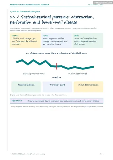
Concept 74
Reading a Belly X-ray
Concept 75
Free Air and Air in the Wrong Places
Concept 76
Twists and Babies' Bellies
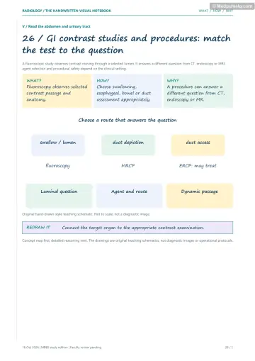
Concept 77
Barium: Painting the Gut
Concept 78
Stomach and Duodenum
Concept 79
Small and Large Bowel Disease
Concept 80
The Acute Abdomen
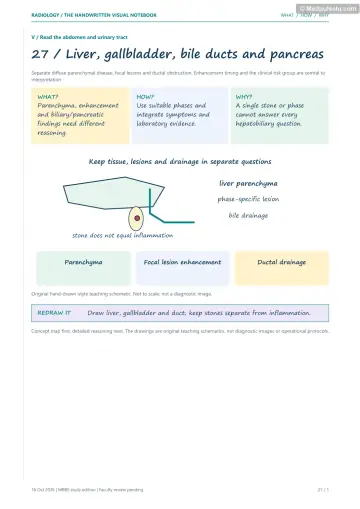
Concept 81
Liver Infections and Cysts
Concept 82
Liver Tumours
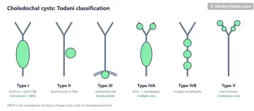
Concept 83
Gallbladder and Bile Ducts
Concept 84
The Pancreas
⚡ Rapid revision
The whole Part on one screen — read it the night before.
Plain films, emergencies and children
- 3‑6‑9 rule (small bowel 3, colon 6, caecum 9 cm). Valvulae conniventes (complete, central) vs haustra (incomplete, peripheral). Step‑ladder = SBO. CT = IOC (cause, closed loop, strangulation).
- SBO: adhesions are common even without prior surgery; also assess hernias and other causes; LBO: colorectal cancer, volvulus. Competent IC valve → caecal perforation risk.
- Free gas: erect CXR best plain film; CT most sensitive. Rigler (double wall), football, falciform ligament, cupola, Doge's cap (Morison's pouch). Chilaiditi = mimic.
- Pneumobilia central (ERCP, anastomosis, gallstone ileus); portal venous gas peripheral (ischaemia, NEC). Rigler's triad: pneumobilia + SBO + ectopic stone.
- Sigmoid volvulus: coffee bean, bent inner tube, Frimann‑Dahl; bird's beak on enema; decompress. Caecal: comma/embryo in LUQ.
- HPS: US ≥ 3 mm thick / ≥ 15 mm long; single bubble. Double bubble = duodenal atresia (Down). Triple = jejunal. Malrotation: upper GI study (DJ flexure low/right), whirlpool on US; bilious vomiting = emergency. Intussusception: US target/pseudokidney; claw sign; air enema reduction. NEC: pneumatosis. Hirschsprung: transition zone, RS index < 1. Meconium ileus: microcolon, soap bubble.
Contrast studies, stomach and bowel
- Leak → water‑soluble contrast. Fistula‑in‑ano IOC = MRI.
- Achalasia: bird's beak, no fundal gas, Hurst phenomenon; manometry gold standard. Carcinoma: shouldered irregular (apple‑core/rat‑tail). DES: corkscrew. Web: anterior post‑cricoid (Plummer–Vinson). Zenker: posterior, Killian's dehiscence. Boerhaave: Naclerio V.
- Benign gastric ulcer: projects outside, Hampton line, radiating folds. Malignant: within lumen, Carman meniscus, clubbed folds. Linitis plastica = leather bottle. Sliding hernia (GEJ up) vs rolling (fundus up, volvulus risk). Reverse‑3 (Frostberg) = pancreatic head mass.
- Crohn's: terminal ileum, skip, string sign of Kantor, cobblestone, comb sign, creeping fat, fistulae; CT/MR enterography. UC: rectum‑up continuous, collar‑stud ulcers, lead‑pipe, toxic megacolon > 6 cm. TB: conical caecum, Fleischner (inverted umbrella), Stierlin, necrotic nodes.
- Diverticulitis: CT. Apple‑core = colorectal cancer; rectal staging MRI. Mexican hat = pedunculated polyp.
- Appendicitis: US (children), CT (adults), MRI (pregnancy); > 6 mm non‑compressible; caecal bar, arrowhead; Alvarado. AMI: CTA (SMA embolus; venous = thick wall); pneumatosis + portal gas late.
Liver, biliary tree and pancreas
- Abscess: US first; CT double target; pyogenic cluster sign, multiple, biliary source; amoebic single right lobe posterosuperior.
- Hydatid (WHO‑IWGE): CE1 sand/double wall; CE2 daughter cysts (rosette); CE3a water‑lily; CE4 ball of wool; CE5 calcified. Lung: water‑lily, Cumbo sign.
- Liver: 75% portal / 25% arterial; HCC arterial. Haemangioma: peripheral nodular centripetal fill, light‑bulb T2. FNH: central scar T2 bright, late enhancement. Fibrolamellar: young, normal AFP, calcified T2‑dark scar. HCC: APHE + washout + capsule (LI‑RADS 5), portal vein tumour thrombus. Mets commonest malignant; hypervascular: NET, RCC, thyroid, melanoma, choriocarcinoma.
- Cirrhosis: caudate hypertrophy, nodular surface, splenomegaly, varices. Surveillance US ± AFP 6‑monthly.
- Gallstones: US, shadowing, mobile; WES sign; 10–15% radio‑opaque; Mercedes‑Benz. Cholecystitis: wall > 3 mm, fluid, sonographic Murphy; HIDA most specific (rim sign = gangrene). MRCP for CBD stones. Adenomyomatosis: comet tails. GB cancer: north India, segments IV/V. Todani I commonest; V = Caroli (central dot).
- Pancreatitis: US for stones; CECT at 72–96 h; Atlanta APFC/pseudocyst (> 4 wk) vs ANC/WON; Balthazar CTSI; sentinel loop, colon cut‑off. Chronic: calcification, chain of lakes; tropical calcific (south India). Cancer: hypoenhancing mass, double duct sign, > 180° SMA encasement unresectable. Serous cystadenoma: central stellate calcified scar.
🎯 Single‑best‑answer MCQs
Pick an answer to see the explanation — and the concept to revisit if you missed it. Your answers are saved on this device.
Score 0 / 00 of 20 answered
Question 1
Dilated centrally placed bowel loops with mucosal folds crossing the entire width of the lumen indicate:
Question 2
The most sensitive investigation for pneumoperitoneum is:
Question 3
Rigler's triad of gallstone ileus consists of:
Question 4
The investigation of choice for suspected hypertrophic pyloric stenosis is:
Question 5
A "double bubble" with no distal bowel gas in a newborn with Down syndrome indicates:
Question 6
A "coffee bean" sign on an abdominal radiograph indicates:
Question 7
A dilated oesophagus with smooth tapering "bird's beak" narrowing at the gastro‑oesophageal junction and absent fundal gas suggests:
Question 8
A posterior outpouching through Killian's dehiscence is a:
Question 9
Which feature on a barium meal favours a benign gastric ulcer?
Question 10
The "string sign of Kantor" is seen in:
Question 11
A conical, shrunken caecum with a thickened gaping ileocaecal valve and narrow terminal ileum (inverted umbrella) suggests:
Question 12
The first imaging investigation for suspected appendicitis in a 9‑year‑old is:
Question 13
The investigation of choice for suspected acute mesenteric ischaemia is:
Question 14
A single abscess in the posterosuperior right lobe of the liver after dysentery is most likely:
Question 15
Floating, detached membranes within a hepatic hydatid cyst produce the:
Question 16
Peripheral nodular discontinuous enhancement with progressive centripetal fill‑in on delayed images is characteristic of:
Question 17
A large liver mass with a calcified, T2‑hypointense central scar in a 22‑year‑old with no cirrhosis and normal AFP is most likely:
Question 18
The most specific imaging test for acute calculous cholecystitis is:
Question 19
The optimal timing of contrast‑enhanced CT to assess severity of acute pancreatitis is:
Question 20
Simultaneous dilatation of the common bile duct and main pancreatic duct suggests:
🃏 Flashcards
Say the answer out loud before you flip. Shuffle once you know the order.
Card 1 of 20
🩺 Staged clinical cases
Read the presentation, decide what is going on and what you would do — then reveal the reasoning.
Case 1 — The vomiting newborn
Stage A: A 2‑day‑old term baby has bilious vomiting. Abdominal radiograph: gas in the stomach and proximal duodenum, with a little distal gas.
Linked concepts: 76. Twists and Babies' Bellies
Case 2 — The old man with a distended abdomen
Stage A: A 75‑year‑old nursing‑home resident with chronic constipation has gross abdominal distension and absolute constipation for 3 days. AXR: massively dilated inverted‑U loop of colon without haustra extending from the pelvis to the right upper quadrant.
Linked concepts: 74. Reading a Belly X-ray, 76. Twists and Babies' Bellies
Case 3 — The cirrhotic with a liver lesion
Stage A: A 61‑year‑old with HBV cirrhosis on 6‑monthly surveillance has a new 2.5 cm lesion on ultrasound. AFP has risen to 340 ng/mL.
Linked concepts: 82. Liver Tumours
Case 4 — Painless jaundice
Stage A: A 66‑year‑old with weight loss has painless jaundice and a palpable, non‑tender gallbladder. US: dilated intra‑ and extrahepatic ducts and a dilated pancreatic duct; no gallstones.
Linked concepts: 84. The Pancreas, 83. Gallbladder and Bile Ducts
🎤 Viva practice
Answer aloud as if an examiner asked you — then check the model points.
Viva 1
How do you differentiate small from large bowel on an abdominal radiograph?
Viva 2
What are the signs of pneumoperitoneum on a supine film?
Viva 3
Distinguish pneumobilia from portal venous gas.
Viva 4
What are the radiological signs of sigmoid volvulus?
Viva 5
Describe the US criteria for HPS.
Viva 6
What are the imaging features of intussusception?
Viva 7
How do you differentiate achalasia from pseudoachalasia?
Viva 8
List the features of a benign gastric ulcer on a barium meal.
Viva 9
How do you distinguish Crohn's disease from ulcerative colitis on imaging?
Viva 10
What are the US criteria for acute appendicitis?
Viva 11
What is the double target sign?
Viva 12
Describe the WHO classification of hydatid cysts.
Viva 13
Why does HCC wash out on delayed images?
Viva 14
What is the Todani classification of choledochal cysts?
Viva 15
Explain the revised Atlanta classification of pancreatic collections.
Keep going
Educational only — not a substitute for clinical judgement, supervision, or local guidelines.