Loading…
Cardiac muscle
Spotter answer: Cardiac muscle with intercalated discs.
Workable field
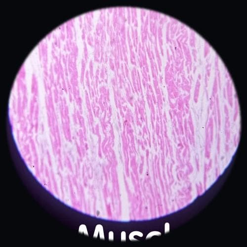
Cardiac muscle at medium power showing longitudinal striated fibres. Cross-striations are not resolvable in this field. Adjust the focus1.0×
- 1Longitudinal striated fibres — Characteristic of cardiac muscle.
Before you name anything
Alright, take a deep breath. Let's start by looking at the overall structure. At low power, notice the arrangement of the fibres. They should appear as branching, which is a key feature of cardiac muscle. Now, focus on the nuclei. They are centrally located, unlike skeletal muscle where they are peripheral. This is your first clue. Next, look for intercalated discs; they might not be very clear, but they are crucial for identifying cardiac muscle. Students often panic by jumping straight to high power and missing these broader features. Avoid that by ensuring you have identified the branching fibres and central nuclei first. Remember, the striations might not be as prominent, but they are there. Keep calm and focus on these distinctive features, and you'll be able to confidently identify this specimen.
Work it out, one rung at a time
Answer each question in your head first. Then open it.
- 1
low-power architecture
What is the arrangement of the fibres?
- 2
epithelium or covering
Where are the nuclei located?
- 3
the signature feature
What unique structure should you find?
- 4
confirm and exclude the look-alike
How do you differentiate from skeletal muscle?
Viva voce
1 of 5 · 0 answeredSay your answer out loud before you open it. That is the exam skill.
What is the function of intercalated discs?