Loading…
🩻 Radiology · Part 8 revision hub
Trauma and Emergency Imaging
Everything you need to revise this Part in one place — the one‑page note for every concept, a rapid‑revision sheet, exam‑style questions that point you back to the right concept, flashcards, staged cases and viva practice.
10 concepts20 MCQs20 flashcards4 cases15 viva questions
📝 The plate wall
Every concept in this Part as a one‑page handwritten note. Open one to read it in full and zoom.
Concept 93
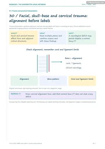
Face and Skull Injuries
Concept 94
The Neck After an Injury
Concept 95
Back and Pelvis Injuries
Concept 96
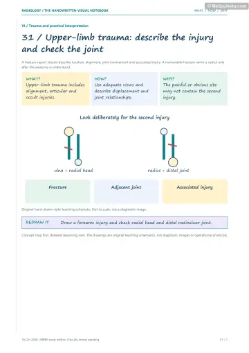
Shoulder and Elbow Injuries
Concept 97
Wrist and Hand Injuries
Concept 98
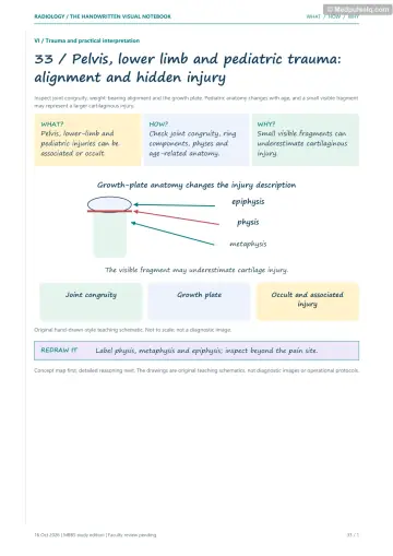
Hip and Knee Injuries
Concept 99
Ankle and Foot Injuries
Concept 100
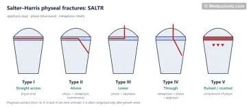
Children's Broken Bones
Concept 101
Bones That Crack from Overuse
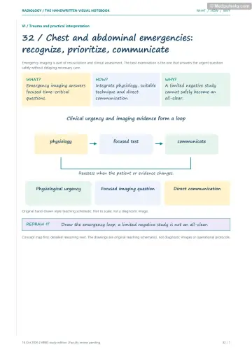
Concept 102
Chest and Belly Injuries
⚡ Rapid revision
The whole Part on one screen — read it the night before.
Head, face and spine
- Face IOC = CT (3D). Blow‑out (floor > medial wall): teardrop (fat/inferior rectus), black eyebrow (orbital emphysema); trapdoor in children. Le Fort (all pterygoid plates): I floating palate, II pyramidal (orbital floor/rim), III craniofacial dysjunction (zygomatic arch). ZMC (tripod/tetrapod). Arch = submentovertex. Mandible: OPG, two fractures.
- C‑spine: NEXUS/Canadian rule; adult IOC CT; MRI for cord/ligaments. ABCS; adequate = C7–T1; four lines; prevertebral C2 ≤ 7 mm, C6 ≤ 22 mm; ADI ≤ 3 mm adult. Pseudosubluxation C2/3 in children (Swischuk line).
- Jefferson: C1 burst (axial load), lateral mass overhang > 7 mm = transverse ligament rupture. Hangman's: bilateral C2 pars (hyperextension; RTA). Odontoid type II commonest, unstable, non‑union. Flexion teardrop = most unstable. Clay‑shoveller's = C7 spinous process, stable. Bilateral facet dislocation > 50% slip. SCIWORA → MRI.
- Thoracolumbar: Denis three columns (middle column key). Compression (osteoporotic, T11–L1). Burst (retropulsion, widened interpedicular distance). Chance (lap belt, horizontal split, bowel/pancreas injury). Calcaneal fracture → check lumbar spine.
- Spondylolysis: collar on Scottie dog; spondylolisthesis: decapitated Scottie dog, inverted Napoleon's hat; Meyerding grades.
- Pelvis = ring (look for second break); Young–Burgess (APC, LC, VS); contrast blush → embolisation. Bladder rupture: CT cystography; intraperitoneal (outlines bowel) → surgery; extraperitoneal (molar tooth) → catheter if uncomplicated; repair selected complicated injuries. Blood at meatus → RGU.
Limbs and children
- Anterior shoulder dislocation (~95%): Hill–Sachs (posterolateral head), Bankart (anteroinferior glenoid). Posterior: light bulb, rim sign, trough line (reverse Hill–Sachs); 3 Es (epilepsy, electric shock, extreme trauma); axillary/Y view. Proximal humerus: surgical neck; Neer. Clavicle: middle third.
- Elbow: sail sign + posterior fat pad = occult intra‑articular fracture (adult radial head; child supracondylar). Anterior humeral line, radiocapitellar line. CRITOE. Supracondylar: AIN, brachial artery, Volkmann, cubitus varus.
- Colles' (dorsal, dinner fork), Smith's (volar), Barton's (intra‑articular rim). MUGR: Monteggia (ulna + radial head), Galeazzi (radius + DRUJ). Scaphoid: commonest carpal, waist, occult → MRI, proximal pole AVN. Terry‑Thomas = SL dissociation. Spilled teacup = lunate dislocation; perilunate = capitate dorsal. Gilula's arcs. Boxer's = 5th MC neck. Bennett's = intra‑articular thumb MC base. Gamekeeper's = thumb UCL (Stener).
- Hip: Shenton's line. Intracapsular (subcapital commonest) → AVN (medial circumflex) → arthroplasty if displaced; Garden I–IV. Intertrochanteric → DHS. Occult → MRI. Posterior dislocation (dashboard; FAdIR; sciatic nerve).
- Knee: lipohaemarthrosis = intra‑articular fracture (tibial plateau); Segond = ACL tear; bipartite patella (superolateral) mimic; MRI for ligaments/menisci.
- Ankle: Ottawa rules; Weber A/B/C (C unstable); trimalleolar; Maisonneuve (proximal fibula). Aviator's astragalus = talar neck; Hawkins sign = good. Calcaneus: Böhler's < 20°, lumbar spine. Lisfranc: 2nd MT base–middle cuneiform alignment, fleck sign. 5th MT: zone 1 pseudo‑Jones (good), zone 2 Jones (non‑union), zone 3 stress. March = 2nd MT.
- Children: torus (buckle), greenstick, plastic bowing, toddler's. Salter–Harris SALTR: II commonest (Thurston‑Holland), V worst. SCFE: Klein's line misses epiphysis, frog‑leg lateral, posteromedial slip. NAI: classic metaphyseal (corner/bucket‑handle) and posterior rib fractures, different ages; skeletal survey < 2 yr. Positioning safety: frog-leg lateral is for stable SCFE; suspected unstable SCFE needs an AP and cross-table lateral without forced hip movement, with urgent orthopaedic review.
- Stress: fatigue (normal bone, abnormal stress) vs insufficiency (weak bone, normal stress); X‑ray late, MRI early; sacral H (Honda) sign; dreaded black line (anterior tibia); high‑risk sites.
Chest and abdominal trauma
- FAST: subxiphoid, Morison's pouch (most sensitive), splenorenal, pelvic; ~200 mL; eFAST adds pneumothorax/haemothorax. Unstable + abdominal FAST positive → urgent laparotomy; pericardial fluid requires the cardiac trauma pathway; stable → CECT.
- Ribs 1–3 = high energy; 10–12 → liver/spleen/kidney. Flail chest ≥ 3 ribs in ≥ 2 places. Contusion within 6 h. Aortic injury at the isthmus: widened mediastinum (> 8 cm), trachea/NG to right, apical cap → CTA. Diaphragmatic rupture: collar sign, dependent viscera sign.
- Spleen = commonest blunt abdominal organ injury; AAST grades; contrast blush → angioembolisation; most non‑operative. Hypoperfusion complex = impending shock.
🎯 Single‑best‑answer MCQs
Pick an answer to see the explanation — and the concept to revisit if you missed it. Your answers are saved on this device.
Score 0 / 00 of 20 answered
Question 1
The "teardrop sign" on coronal CT of the orbit indicates:
Question 2
A Le Fort fracture that separates the entire facial skeleton from the cranium through the frontozygomatic sutures and zygomatic arches is:
Question 3
On a lateral cervical spine radiograph, the film is adequate only if it shows:
Question 4
Bilateral fractures through the pars interarticularis of C2 constitute:
Question 5
The commonest and most unstable type of odontoid fracture with the highest non‑union rate is:
Question 6
A horizontal fracture through the spinous process, pedicles and vertebral body after a lap‑belt injury is a:
Question 7
On CT cystography, contrast outlining bowel loops and filling the paracolic gutters indicates:
Question 8
After an epileptic seizure, a patient's AP shoulder radiograph shows a symmetrically rounded humeral head ("light bulb"). The diagnosis is:
Question 9
An elevated anterior fat pad ("sail sign") and visible posterior fat pad after elbow trauma in an adult most often indicate an occult fracture of the:
Question 10
A fracture of the distal third of the radius with dislocation of the distal radioulnar joint is a:
Question 11
A widened scapholunate gap (> 3 mm) on a PA wrist radiograph is called the:
Question 12
The investigation of choice for a suspected hip fracture when radiographs are normal is:
Question 13
A small avulsion fracture of the lateral tibial rim (Segond fracture) is strongly associated with:
Question 14
A fracture of the 5th metatarsal at the metaphyseal–diaphyseal junction with a high non‑union risk is:
Question 15
After a fall from height with a calcaneal fracture, which additional region must be imaged?
Question 16
The commonest Salter–Harris physeal fracture is:
Question 17
In slipped capital femoral epiphysis, Klein's line drawn along the superior femoral neck on the AP view:
Question 18
Which fracture is most specific for non‑accidental injury in an infant?
Question 19
The most sensitive early imaging test for a stress fracture is:
Question 20
A haemodynamically unstable trauma patient has free fluid in Morison's pouch on FAST. The next step is:
🃏 Flashcards
Say the answer out loud before you flip. Shuffle once you know the order.
Card 1 of 20
🩺 Staged clinical cases
Read the presentation, decide what is going on and what you would do — then reveal the reasoning.
Case 1 — The diver
Stage A: A 19‑year‑old dives into a shallow pond and strikes his head. He has neck pain but moves all limbs. He is immobilised.
Linked concepts: 94. The Neck After an Injury
Case 2 — The fall on the outstretched hand
Stage A: A 24‑year‑old falls while cycling; she has anatomical snuffbox tenderness. Scaphoid series radiographs are normal.
Linked concepts: 97. Wrist and Hand Injuries
Case 3 — The elderly woman who cannot stand
Stage A: An 84‑year‑old falls in the bathroom and cannot bear weight. Her leg is shortened and externally rotated. AP pelvis: subtle cortical step at the subcapital region? Shenton's line looks intact.
Linked concepts: 98. Hip and Knee Injuries
Case 4 — The unstable motorcyclist
Stage A: A 28‑year‑old motorcyclist arrives with BP 80/50 mmHg, HR 135/min despite 1 L crystalloid. eFAST shows free fluid in Morison's pouch and the splenorenal recess; lung sliding present bilaterally. Pelvic radiograph: open‑book symphyseal diastasis of 4 cm.
Linked concepts: 102. Chest and Belly Injuries, 95. Back and Pelvis Injuries
🎤 Viva practice
Answer aloud as if an examiner asked you — then check the model points.
Viva 1
What are the four lines on a lateral cervical spine radiograph?
Viva 2
Classify odontoid fractures.
Viva 3
Describe the Le Fort fractures.
Viva 4
What is a Chance fracture and why does it matter?
Viva 5
How do you differentiate intraperitoneal from extraperitoneal bladder rupture?
Viva 6
What lesions accompany anterior shoulder dislocation?
Viva 7
What is the significance of the elbow fat pad sign?
Viva 8
Why is the scaphoid prone to AVN?
Viva 9
Differentiate intracapsular from extracapsular femoral fractures.
Viva 10
What is the Weber classification?
Viva 11
What is a Maisonneuve fracture?
Viva 12
What is the Hawkins sign?
Viva 13
List the Salter–Harris types.
Viva 14
What skeletal findings suggest non‑accidental injury?
Viva 15
What are the four FAST windows and the management algorithm?
Keep going
Educational only — not a substitute for clinical judgement, supervision, or local guidelines.