Loading…
🩻 Radiology · Part 4 revision hub
Musculoskeletal Imaging
Everything you need to revise this Part in one place — the one‑page note for every concept, a rapid‑revision sheet, exam‑style questions that point you back to the right concept, flashcards, staged cases and viva practice.
16 concepts20 MCQs20 flashcards4 cases15 viva questions
📝 The plate wall
Every concept in this Part as a one‑page handwritten note. Open one to read it in full and zoom.
Concept 43
How to Read a Bone X-ray
Concept 44
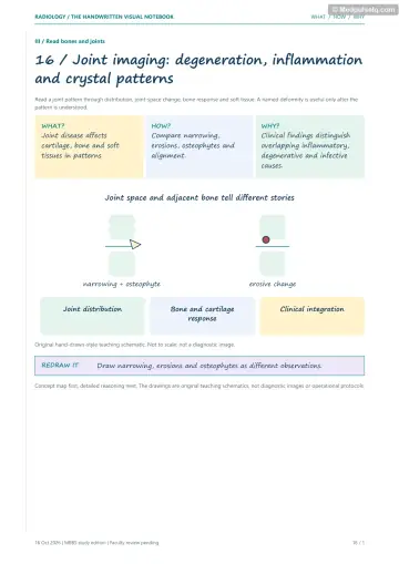
Worn-out versus Inflamed Joints
Concept 45
Rheumatoid Arthritis
Concept 46
The Spine That Turns to Bamboo
Concept 47
Crystals in Joints
Concept 48
Joints That Cannot Feel and Spines That Ossify

Concept 49
Infected Bone
Concept 50
Infected Joints, TB Bones and the Diabetic Foot
Concept 51
Soft Bones from Missing Vitamins
Concept 52
Hormones, Blood and Bone
Concept 53
Bone That Dies
Concept 54
Reading a Bone Lump
Concept 55
Friendly Bone Lumps
Concept 56
Dangerous Bone Tumours
Concept 57
Cancer That Spreads to Bone

Concept 58
Bones Built Differently
⚡ Rapid revision
The whole Part on one screen — read it the night before.
Fractures and arthritis
- Two orthogonal views, joint above and below; ABCS (alignment, bone, cartilage, soft tissue). Describe the distal fragment relative to the proximal. Lipohaemarthrosis = intra‑articular fracture.
- OA = LOSS (non‑uniform loss, osteophytes, sclerosis, cysts); medial knee earliest. Inflammatory = uniform loss, periarticular osteopenia, marginal erosions.
- RA: symmetric MCP/PIP/wrist, DIP spared, ulnar styloid and 5th MTP erosions; swan neck, boutonnière, Z‑thumb, ulnar deviation; atlantoaxial subluxation. Felty = RA + spleen + neutropenia; Caplan = RA + pneumoconiosis nodules.
- AS: bilateral symmetric sacroiliitis (MRI earliest), Romanus shiny corner, squaring, syndesmophytes → bamboo spine; dagger, trolley‑track; carrot‑stick fracture.
- Psoriatic: DIP, sausage digit, pencil‑in‑cup, ivory phalanx, no osteopenia, parasyndesmophytes.
- Gout: 1st MTP, punched‑out erosions with overhanging edges (Martel), joint space preserved, no osteopenia; DECT. CPPD: meniscal chondrocalcinosis, isolated patellofemoral OA; 4 Hs. Ochronosis: multilevel disc calcification.
- Charcot 6 Ds: diabetes → foot (Lisfranc), syringomyelia → shoulder, tabes → knee. DISH ≥ 4 vertebrae, candle wax, discs and SIJ normal. OPLL cervical, MRI for cord.
Infection and metabolic disease
- Osteomyelitis: radiograph lags ~10–14 days; bone scan 24–48 h; MRI IOC. Sequestrum (dead), involucrum (new shell), cloaca (hole); Marjolin ulcer. Brodie abscess: subacute metaphyseal lucency with sclerotic rim, penumbra sign.
- Septic arthritis: US effusion → aspirate; early widening → rapid narrowing; Tom Smith (infant hip). TB arthritis: Phemister triad; spina ventosa; Pott's spine (paradiscal, disc preserved early, large cold abscess).
- Diabetic foot osteomyelitis: MRI (low T1 marrow contiguous with ulcer).
- Rickets: widened physis, cupping, splaying, fraying (wrist, knee); healing dense band. Osteomalacia: Looser zones. Osteoporosis: DEXA T ≤ −2.5. Scurvy: Wimberger ring, Frankel line, Trümmerfeld zone, Pelkan spur, subperiosteal haemorrhage.
- HPT: radial middle phalanges (2nd/3rd) subperiosteal resorption, salt‑and‑pepper skull, brown tumours. Renal: rugger‑jersey. Paget's: enlarged bone, cotton‑wool skull, picture‑frame vertebra, blade of grass. Thalassaemia: hair‑on‑end, Erlenmeyer flask; sickle: H vertebra, AVN.
- AVN: steroids/alcohol; femoral head; MRI (double line sign); crescent sign = collapse. Hawkins sign = viable talus. Perthes 4–8 yr boys, Waldenström sign earliest. Kienböck lunate, Freiberg 2nd MT head, Köhler navicular, Osgood–Schlatter tibial tuberosity.
Tumours and dysplasias
- ALTA: Age (most useful), Location, Transition zone, Appearance (matrix, periosteum). Epiphysis: chondroblastoma (immature), GCT (mature). Diaphysis: Ewing. Metaphysis: osteosarcoma.
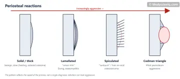
- Periosteal reactions: solid (benign) → lamellated (Ewing, infection) → sunburst (osteosarcoma) → Codman triangle (aggressive).
- Polyostotic: FEEMMHI. "Don't touch": NOF, bone island, cortical desmoid.
- Osteochondroma (commonest benign; points away from joint; cap > 1.5–2 cm = worry). Osteoid osteoma (nidus < 1.5 cm, night pain, NSAIDs, CT, RFA). SBC (proximal humerus, fallen fragment). ABC (fluid–fluid levels). FD (ground glass, shepherd's crook, McCune–Albright). Chondroblastoma (epiphyseal, chicken wire). Haemangioma (corduroy, polka dot).
- Osteosarcoma (10–25, distal femur, sunburst, Codman, lung mets). Parosteal (posterior distal femur, string sign). Ewing (5–20, diaphysis, onion skin, soft‑tissue mass). Chondrosarcoma (> 40, pelvis, rings and arcs). GCT (20–40, epimetaphyseal to subchondral, soap bubble, no sclerotic rim). Chordoma (sacrum > clivus).
- Metastasis = commonest malignant bone lesion; prostate sclerotic; renal/thyroid blow‑out; winking owl. Myeloma: raindrop skull, cold bone scan, WB‑LDCT/MRI. LCH: bevelled edge, vertebra plana (child).
- Achondroplasia: champagne‑glass pelvis, trident hand, narrowing interpedicular distance. OI: fractures, Wormian bones, blue sclera. Osteopetrosis: bone‑within‑bone, sandwich. Cleidocranial: absent clavicles. Morquio central beak; Hurler inferior beak.
🎯 Single‑best‑answer MCQs
Pick an answer to see the explanation — and the concept to revisit if you missed it. Your answers are saved on this device.
Score 0 / 00 of 20 answered
Question 1
A horizontal‑beam lateral knee radiograph after trauma shows a fat–fluid level in the suprapatellar bursa. This indicates:
Question 2
Which feature favours osteoarthritis over rheumatoid arthritis?
Question 3
Which joints are characteristically spared in rheumatoid arthritis?
Question 4
The earliest imaging finding of ankylosing spondylitis is best shown by:
Question 5
Punched‑out juxta‑articular erosions with overhanging edges and preserved joint space at the first MTP joint suggest:
Question 6
A neuropathic shoulder joint is most characteristically associated with:
Question 7
Flowing ossification along the anterolateral aspect of at least four contiguous thoracic vertebrae with preserved disc heights and normal SI joints is:
Question 8
The investigation of choice for early acute osteomyelitis is:
Question 9
In chronic osteomyelitis, the dead fragment of bone acting as a nidus of infection is the:
Question 10
TB dactylitis of the short tubular bones of a child's hand is called:
Question 11
Looser zones are characteristic of:
Question 12
The radiographic hallmark of rickets at the wrist is:
Question 13
The earliest and most specific radiographic sign of hyperparathyroidism is subperiosteal resorption of:
Question 14
H‑shaped vertebrae are characteristic of:
Question 15
The most sensitive imaging test for early avascular necrosis of the femoral head is:
Question 16
Which tumour classically produces an onion‑skin periosteal reaction in the diaphysis of a child's long bone?
Question 17
A "fallen fragment" sign is pathognomonic of:
Question 18
A lytic, eccentric, expansile lesion extending to the subchondral bone of the distal femur in a 28‑year‑old, with a narrow zone of transition and no sclerotic rim, is most likely:
Question 19
A bone scan is often normal despite extensive disease in:
Question 20
A "champagne‑glass" pelvic inlet and trident hand are features of:
🃏 Flashcards
Say the answer out loud before you flip. Shuffle once you know the order.
Card 1 of 20
🩺 Staged clinical cases
Read the presentation, decide what is going on and what you would do — then reveal the reasoning.
Case 1 — The limping 6‑year‑old
Stage A: A 6‑year‑old boy has had a painless limp for 3 weeks and intermittent knee pain. He is afebrile with normal inflammatory markers. AP pelvis: subtle widening of the left medial hip joint space and a slightly smaller, denser left femoral epiphysis.
Linked concepts: 53. Bone That Dies, 50. Infected Joints, TB Bones and the Diabetic Foot
Case 2 — The teenager's night pain
Stage A: A 15‑year‑old has 2 months of left knee pain waking him at night, not relieved by NSAIDs, and a firm swelling above the knee. Radiograph: ill‑defined mixed lytic–sclerotic distal femoral metaphyseal lesion with spiculated periosteal reaction and a Codman triangle.
Linked concepts: 54. Reading a Bone Lump, 56. Dangerous Bone Tumours
Case 3 — The diabetic's swollen foot
Stage A: A 58‑year‑old with long‑standing diabetes and peripheral neuropathy has a warm, swollen, minimally painful midfoot. No ulcer. Radiograph: fragmentation and subluxation at the tarsometatarsal joints with sclerosis.
Case 4 — The old man with back pain and anaemia
Stage A: A 68‑year‑old has back pain, anaemia, raised creatinine and calcium. Spine radiograph: diffuse osteopenia with an L2 compression fracture. A bone scan done elsewhere was "normal".
Linked concepts: 57. Cancer That Spreads to Bone, 22. Concept 22
🎤 Viva practice
Answer aloud as if an examiner asked you — then check the model points.
Viva 1
How do you describe a fracture?
Viva 2
Distinguish OA from RA radiographically.
Viva 3
What is the significance of atlantoaxial subluxation in RA?
Viva 4
Describe the spinal changes of AS.
Viva 5
What are the radiological features of chronic gout?
Viva 6
List the 6 Ds of a Charcot joint and causes by site.
Viva 7
Why are radiographs normal in early osteomyelitis?
Viva 8
Define sequestrum, involucrum and cloaca.
Viva 9
How do you differentiate osteoporosis from osteomalacia?
Viva 10
What are the radiological signs of scurvy?
Viva 11
Describe the MRI features of AVN.
Viva 12
What does ALTA stand for in bone tumour assessment?
Viva 13
Name four "don't touch" bone lesions.
Viva 14
Compare osteosarcoma and Ewing sarcoma.
Viva 15
Why is a bone scan unreliable in myeloma?
Keep going
Educational only — not a substitute for clinical judgement, supervision, or local guidelines.