Loading…
🩻 Radiology · Part 5 revision hub
Neuroradiology
Everything you need to revise this Part in one place — the one‑page note for every concept, a rapid‑revision sheet, exam‑style questions that point you back to the right concept, flashcards, staged cases and viva practice.
15 concepts20 MCQs20 flashcards4 cases15 viva questions
📝 The plate wall
Every concept in this Part as a one‑page handwritten note. Open one to read it in full and zoom.
Concept 59
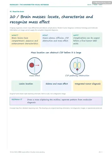
Reading a Brain Scan
Concept 60
Tumours Outside the Brain
Concept 61
Tumours Inside the Brain
Concept 62
Tumours Around the Pituitary and Ventricles
Concept 63
Skin-and-Brain Syndromes
Concept 64
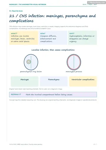
Brain Infections I
Concept 65
Brain Infections II
Concept 66
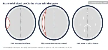
Head Injury: Where Is the Blood?
Concept 67
Hidden Head Injuries
Concept 68
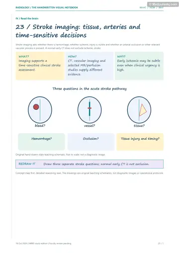
Stroke: Time Is Brain
Concept 69
Other Kinds of Stroke
Concept 70
Bleeding Inside the Brain
Concept 71
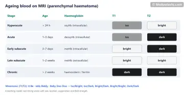
How Blood Changes on MRI
Concept 72
Babies' Brains and the Brain's Wiring
Concept 73
The Spine and Spinal Cord
⚡ Rapid revision
The whole Part on one screen — read it the night before.
Approach and tumours
- CT (~1 min): trauma, stroke, SAH, status epilepticus. MRI (30–40 min): tumour, epilepsy, MS, DAI, posterior fossa.
- Extra‑axial: broad dural base, CSF cleft, grey matter buckling, hyperostosis, dural tail. Intra‑axial: within parenchyma.
- Meningioma: commonest primary intracranial tumour; hyperdense, calcified, intense enhancement, dural tail, sunburst/mother‑in‑law sign, alanine peak.
- CPA: vestibular schwannoma (ice‑cream cone, widened IAC) > meningioma > epidermoid (DWI bright). Arachnoid cyst = CSF everywhere.
- Adult: metastasis commonest (grey–white junction, multiple); GBM commonest primary malignant (thick irregular ring, necrosis, butterfly). Oligodendroglioma: frontal, calcified. Lymphoma: periventricular, hyperdense, restricts.
- Child posterior fossa: pilocytic (hemisphere, cyst + nodule, best prognosis), medulloblastoma (vermis, hyperdense, restricts, drop mets), ependymoma (4th ventricle, plastic, calcified), DIPG (pons). Adult cyst + nodule = haemangioblastoma.
- Microadenoma: dynamic MRI (enhances later). Macroadenoma: snowman, bitemporal hemianopia. Craniopharyngioma: child, cystic + calcified + solid. Colloid cyst: anterior 3rd ventricle, hyperdense, sudden death. Retinoblastoma: calcified, MRI.
- NF1 optic glioma, bare orbit; NF2 bilateral schwannomas (MISME); TS tubers, calcified subependymal nodules, SEGA, renal AML; VHL haemangioblastoma + RCC; Sturge–Weber port‑wine + tram‑track calcification.
Infection and trauma
- Meningitis = LP; imaging for contraindications/complications. TBM: basal exudates, hydrocephalus, basal ganglia infarcts; tuberculoma T2 dark, target.
- Abscess: smooth thin ring, thinner medial wall, central DWI restriction. Ring lesions: MAGIC DR + TB + NCC; open ring = demyelination.
- NCC: vesicular (hole with dot) → colloidal (ring + oedema) → granular nodular → calcified. Commonest acquired epilepsy cause in India. Racemose = basal cisterns, no scolex.
- HSV: medial temporal + insula, basal ganglia spared; acyclovir now. JE: bilateral thalami. HIV: toxo (multiple basal ganglia rings, eccentric target), lymphoma (single, restricts, thallium‑hot), PML (U‑fibres, no enhancement).
- EDH: biconvex, MMA, doesn't cross sutures, lucid interval. SDH: crescentic, bridging veins, crosses sutures not falx. tSAH: commonest traumatic bleed. Contusion: frontal/temporal poles. Blood: hyper → iso (1–3 wk) → hypo.
- DAI: MRI SWI; grade 1 grey–white, 2 corpus callosum (splenium), 3 brainstem. Mount Fuji = tension pneumocephalus. Growing skull fracture < 3 yr. Uncal herniation → CN III palsy.
Vascular, blood, neonatal and spine
- Stroke: NCCT first; hyperdense MCA, insular ribbon, lentiform obscuration; ASPECTS. DWI earliest. Peak oedema days 3–5; fogging 2–3 weeks. IV lysis ≤ 4.5 h (tenecteplase = alteplase); thrombectomy ≤ 24 h with selection, large core eligible (2026).
- Lacune < 15 mm deep. CVT: cord sign, dense triangle, empty delta (CECT), non‑arterial haemorrhagic infarcts, MRV/CTV. Hypoxic: reversal and white cerebellum signs. Moyamoya: puff of smoke (DSA), ivy sign. PRES: parieto‑occipital, eclampsia, tacrolimus, reversible.
- ICH: putamen = hypertension; lobar elderly = amyloid; swirl/spot = expanding. SAH: berry aneurysm (ACom), NCCT then CTA, DSA gold standard; vasospasm days 4–14. AVM: bag of worms; cavernoma: popcorn + haemosiderin rim, angio‑occult; vein of Galen: neonatal heart failure.
- Blood on MRI (T1/T2): IB, ID, BD, BB, DD; metHb = T1 bright; haemosiderin = dark, blooms.
- Preterm: cranial US (IVH grades I–IV, PVL cysts). Term profound HIE: thalami, putamina, perirolandic. NPH: wet, wacky, wobbly; Evans > 0.3, DESH. MS: Dawson's fingers, open ring, McDonald DIS/DIT. Trident pons = osmotic demyelination.
- Spine MRI IOC. L4–5/L5–S1 commonest. Extrusion > base. Cauda equina = emergency MRI. Pott's: T10–L2, paradiscal, disc preserved early, subligamentous skip, large calcified cold abscess, gibbus. Intramedullary: ependymoma (adult), astrocytoma (child).
🎯 Single‑best‑answer MCQs
Pick an answer to see the explanation — and the concept to revisit if you missed it. Your answers are saved on this device.
Score 0 / 00 of 20 answered
Question 1
Which feature indicates an extra‑axial location of an intracranial mass?
Question 2
The commonest mass at the cerebellopontine angle is:
Question 3
A CPA lesion that follows CSF on T1 and T2 but is bright on DWI is most likely:
Question 4
A cerebellar hemispheric cystic lesion with an enhancing mural nodule in a 7‑year‑old is most likely:
Question 5
Which paediatric posterior fossa tumour is typically hyperdense on CT, midline, restricts diffusion and seeds the CSF?
Question 6
A calcified, cystic and solid suprasellar mass in a 9‑year‑old is most likely:
Question 7
A hyperdense, non‑enhancing round lesion at the foramen of Monro causing positional headaches is a:
Question 8
Bilateral vestibular schwannomas are diagnostic of:
Question 9
The imaging feature that best distinguishes a pyogenic brain abscess from a necrotic glioblastoma is:
Question 10
The triad of basal meningeal enhancement, hydrocephalus and basal ganglia infarcts suggests:
Question 11
A cyst with an eccentric dot and no surrounding oedema in a young Indian with seizures represents which stage of neurocysticercosis?
Question 12
In an AIDS patient, multiple ring‑enhancing lesions in the basal ganglia with an eccentric target sign suggest:
Question 13
A biconvex hyperdense extra‑axial collection that does not cross suture lines is a:
Question 14
The investigation of choice for suspected diffuse axonal injury is:
Question 15
The earliest imaging modality to detect an acute ischaemic infarct is:
Question 16
The "fogging effect" in cerebral infarction on CT typically occurs at:
Question 17
An empty delta sign on contrast‑enhanced CT indicates:
Question 18
The commonest site of hypertensive intracerebral haemorrhage is the:
Question 19
A haematoma that is hyperintense on both T1 and T2 is in which stage?
Question 20
Which feature favours tuberculous over pyogenic spondylodiscitis?
🃏 Flashcards
Say the answer out loud before you flip. Shuffle once you know the order.
Card 1 of 20
🩺 Staged clinical cases
Read the presentation, decide what is going on and what you would do — then reveal the reasoning.
Case 1 — The thunderclap headache
Stage A: A 48‑year‑old hypertensive woman has a sudden severe occipital headache "like being hit with a bat", followed by vomiting and neck stiffness. GCS 15. NCCT at 3 hours shows hyperdensity in the interhemispheric fissure and suprasellar cistern.
Linked concepts: 70. Bleeding Inside the Brain, 66. Head Injury: Where Is the Blood?
Case 2 — The talking then drowsy boy
Stage A: A 16‑year‑old is struck on the left temple by a cricket ball, loses consciousness briefly, then talks normally. Two hours later he is drowsy with a dilating left pupil.
Linked concepts: 66. Head Injury: Where Is the Blood?, 67. Hidden Head Injuries
Case 3 — The young woman with a first seizure
Stage A: A 21‑year‑old student from a rural district has a first generalised seizure. CT: a 12 mm ring‑enhancing lesion in the right parietal lobe with surrounding oedema and an eccentric nodular dot; two small calcified foci elsewhere.
Linked concepts: 65. Brain Infections II, 64. Brain Infections I
Case 4 — The wake‑up stroke
Stage A: A 72‑year‑old wakes at 6 a.m. with right hemiparesis and aphasia; last seen well at 11 p.m. NCCT: no haemorrhage, ASPECTS 8, hyperdense left M1.
Linked concepts: 68. Stroke: Time Is Brain
🎤 Viva practice
Answer aloud as if an examiner asked you — then check the model points.
Viva 1
How do you differentiate intra‑ from extra‑axial masses?
Viva 2
What are the imaging features of meningioma?
Viva 3
Name the commonest posterior fossa tumours of childhood and their features.
Viva 4
What is the "snowman" sign?
Viva 5
Describe the features of each neurocutaneous syndrome.
Viva 6
What is the role of imaging in meningitis?
Viva 7
List the stages of neurocysticercosis.
Viva 8
Where does HSV encephalitis localise?
Viva 9
Differentiate EDH from SDH.
Viva 10
What is diffuse axonal injury and how is it graded?
Viva 11
List the early CT signs of MCA infarction.
Viva 12
What are the current treatment windows for acute ischaemic stroke?
Viva 13
What are the imaging signs of cerebral venous thrombosis?
Viva 14
Explain the evolution of haematoma signal on MRI.
Viva 15
How do you differentiate pyogenic from tuberculous spondylodiscitis?
Keep going
Educational only — not a substitute for clinical judgement, supervision, or local guidelines.