Loading…
🩻 Radiology · Part 3 revision hub
Chest Imaging
Everything you need to revise this Part in one place — the one‑page note for every concept, a rapid‑revision sheet, exam‑style questions that point you back to the right concept, flashcards, staged cases and viva practice.
17 concepts20 MCQs20 flashcards4 cases15 viva questions
📝 The plate wall
Every concept in this Part as a one‑page handwritten note. Open one to read it in full and zoom.
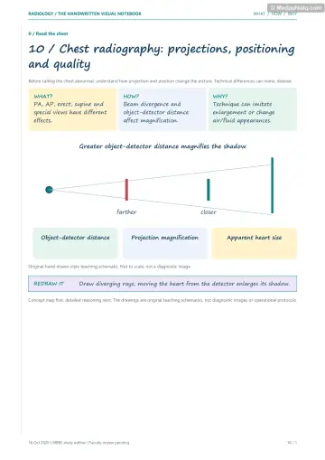
Concept 26
How a Chest X-ray Is Taken
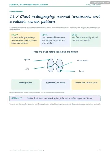
Concept 27
Is This a Good Film, and What Is Normal?
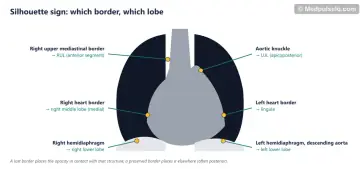
Concept 28
The Silhouette Sign
Concept 29
Fluffy or Lacy? Lung Patterns
Concept 30
Pneumonia, Abscess and Fungus Balls
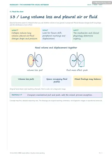
Concept 31
When a Lobe Collapses
Concept 32
Fluid Around the Lung
Concept 33
Air Where It Should Not Be
Concept 34
The Heart on a Chest X-ray
Concept 35
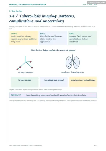
Tuberculosis I: First Infection and Return
Concept 36
Tuberculosis II: Miliary TB and Its Scars
Concept 37
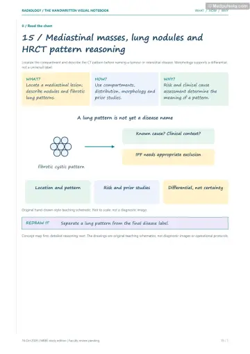
Lumps in the Middle of the Chest
Concept 38
Lung Cancer and the Lonely Nodule
Concept 39
HRCT: Looking Closer at Lung Tissue
Concept 40
Clots in the Lung
Concept 41
Tubes, Lines and Swallowed Things
Concept 42
Little Chests
⚡ Rapid revision
The whole Part on one screen — read it the night before.
Technique and normal anatomy
- PA erect, centred at T7, full inspiration, SID ~180 cm = standard. AP magnifies the heart (don't apply CTR < 0.5).
- Effusion detection: decubitus ~5–25 mL < lateral ~75 mL < PA ~200 mL; US is IOC. Lordotic view: apices, RML. Expiratory film: FB air trapping.
- RIPE: rotation (clavicles equidistant from spinous processes), inspiration (6 anterior / 10 posterior ribs), penetration (vertebrae just visible through heart), exposure/field.
- CTR = (A₁ + A₂)/B; < 0.5 normal on PA. Left hilum higher than right (~97%). Review areas: apices, hila, retrocardiac, below the diaphragm, bones.
- Silhouette sign: right heart border → RML; left heart border → lingula; aortic knuckle → LUL; right dome → RLL; left dome/descending aorta → LLL. Air bronchogram = airspace disease.
Lung patterns, infection, collapse and pleura
- Airspace: fluffy, confluent, air bronchograms; 5 Ps (pus, pulmonary oedema, haemorrhage, proteinaceous, primary cells). Interstitial: lines, dots; Kerley B lines.
- Pneumatocele → Staph; bulging fissure → Klebsiella; perihilar GGO in HIV → PCP. Aspiration supine → posterior RUL / superior RLL.
- Abscess: spherical, thick wall, acute angle, IOC CECT. Empyema: lenticular, obtuse angle, split pleura sign.
- Aspergilloma: Monod sign. ABPA: central bronchiectasis, finger‑in‑glove. Angioinvasive: halo → air crescent.
- Collapse: fissure displacement (most reliable). RUL: Golden S (hilar mass). LUL: veil + Luftsichel. RML: lost right heart border. RLL: superior triangle. LLL: flat waist, retrocardiac sail.
- Opaque hemithorax: collapse pulls, effusion pushes, consolidation central.
- Effusion: CP blunting (~200 mL), meniscus; subpulmonic (lateral peak); phantom tumour (CHF). TB = commonest exudate in young Indian adults.
- Pneumothorax: visceral pleural line; supine deep sulcus sign; USG: no sliding, barcode, lung point. Tension: decompress first. Hydropneumothorax: straight air‑fluid level. Naclerio V = oesophageal rupture. Holly leaf = calcified asbestos plaque.
Heart, TB, masses, ILD, PE and paediatrics
- PVH: upper lobe diversion → Kerley B lines/cuffing → bat‑wing → effusions. Boot = TOF; egg on string = TGA; snowman = TAPVC; box = Ebstein's; water bottle = pericardial effusion; rib notching + 3 sign = coarctation.
- Primary TB: ipsilateral hilar/paratracheal nodes (rim‑enhancing), any lobe, effusion. Post‑primary: apical/posterior upper lobes + superior lower lobe, cavitation, tree‑in‑bud.
- Miliary 1–3 mm random nodules, IOC HRCT. Ranke = calcified Ghon complex; Simon = apical haematogenous; Assmann = infraclavicular; Weigert = vein wall; Rasmussen = PA aneurysm in cavity.
- Mediastinum: anterior 4 Ts (thymoma commonest adult); middle: nodes (adults), duplication cysts (children); posterior: neurogenic.
- Adeno: peripheral, commonest, women/non‑smokers. Squamous: central, cavitates, ↑Ca. SCLC: central, SVCO. Popcorn = hamartoma. Cannonball = RCC, choriocarcinoma. Fleischner 2017: < 6 mm low risk, no follow‑up.
- UIP: subpleural basal honeycombing; NSIP: GGO with subpleural sparing. Signet ring = bronchiectasis. Silicosis: upper zone, eggshell nodes, TB risk. Asbestosis: lower zone + plaques.
- PE: CTPA IOC; polo mint; Hampton hump, Westermark, Fleischner, Palla. V/Q in pregnancy, CKD, allergy, CTEPH.
- ETT 5 cm above carina. Peanut = radiolucent → air trapping. Oesophageal coin en face on AP. Button battery = emergency.
- Neonate: RDS (premature, low volume, ground glass, air bronchograms); TTN (caesarean, fissural fluid, clears 24–72 h); MAS (post‑term, hyperinflation, coarse); Bochdalek CDH (left). Thymic sail = normal.
🎯 Single‑best‑answer MCQs
Pick an answer to see the explanation — and the concept to revisit if you missed it. Your answers are saved on this device.
Score 0 / 00 of 20 answered
Question 1
The most sensitive plain radiographic view for a small pleural effusion is:
Question 2
On a PA CXR, adequate inspiration is indicated by:
Question 3
Loss of the left heart border with a preserved left hemidiaphragm localises disease to the:
Question 4
Air bronchograms are a feature of:
Question 5
A thin‑walled cystic lesion developing during staphylococcal pneumonia in a child is a:
Question 6
Which favours empyema over lung abscess on CT?
Question 7
The Golden S sign indicates:
Question 8
The Luftsichel sign is seen in:
Question 9
The earliest sign of pleural effusion on an erect PA radiograph is:
Question 10
On a supine trauma radiograph, the sign suggesting pneumothorax is:
Question 11
The most specific ultrasound sign of pneumothorax is:
Question 12
The earliest radiographic sign of pulmonary venous hypertension is:
Question 13
A boot‑shaped heart with oligaemic lung fields suggests:
Question 14
The hallmark radiological feature of primary pulmonary tuberculosis in children is:
Question 15
A calcified Ghon complex is called:
Question 16
The commonest posterior mediastinal mass is:
Question 17
"Popcorn" calcification in a solitary pulmonary nodule suggests:
Question 18
On HRCT, a bronchus larger than its accompanying pulmonary artery (signet ring sign) indicates:
Question 19
The investigation of choice in a haemodynamically stable patient with suspected PE and normal renal function is:
Question 20
A term baby born by elective caesarean has tachypnoea, streaky perihilar markings and fluid in the fissures that clear within 48 hours. The diagnosis is:
🃏 Flashcards
Say the answer out loud before you flip. Shuffle once you know the order.
Card 1 of 20
🩺 Staged clinical cases
Read the presentation, decide what is going on and what you would do — then reveal the reasoning.
Case 1 — The silent right heart border
Stage A: A 58‑year‑old smoker has had a cough for 6 weeks despite two antibiotic courses. PA CXR: hazy right paracardiac opacity with loss of the right heart border; right hemidiaphragm crisp; horizontal fissure displaced downward.
Linked concepts: 28. The Silhouette Sign, 31. When a Lobe Collapses
Case 2 — The breathless young man
Stage A: A 21‑year‑old tall, thin man has sudden left chest pain and breathlessness. SpO₂ 95%, BP normal. Erect CXR: visceral pleural line 2.5 cm from the chest wall at the hilum level.
Linked concepts: 33. Air Where It Should Not Be
Case 3 — The stone cutter's cough
Stage A: A 42‑year‑old sandstone mine worker from Rajasthan has progressive breathlessness. CXR: multiple small nodules in both upper zones and hilar nodes with rim calcification.
Case 4 — The ICU film after intubation
Stage A: After emergency intubation, a patient's SpO₂ falls and breath sounds are reduced on the left. CXR: ETT tip below the carina in the right main bronchus; left lung opacified with leftward mediastinal shift.
Linked concepts: 41. Tubes, Lines and Swallowed Things, 31. When a Lobe Collapses
🎤 Viva practice
Answer aloud as if an examiner asked you — then check the model points.
Viva 1
Why is the PA view preferred to the AP view?
Viva 2
How do you assess rotation on a CXR?
Viva 3
Explain the silhouette sign with two examples.
Viva 4
List the 5 Ps of airspace opacification.
Viva 5
How do you differentiate lung abscess from empyema on CT?
Viva 6
Describe the radiographic signs of LUL collapse.
Viva 7
What are the causes of an opaque hemithorax with a central trachea?
Viva 8
What are the ultrasound signs of pneumothorax?
Viva 9
Stage pulmonary venous hypertension on CXR.
Viva 10
Differentiate primary from post‑primary TB on imaging.
Viva 11
List causes of a miliary pattern.
Viva 12
What are the "4 Ts" of the anterior mediastinum?
Viva 13
Name the features of a benign solitary pulmonary nodule.
Viva 14
Differentiate UIP from NSIP on HRCT.
Viva 15
How do you confirm nasogastric tube position on CXR?
Keep going
Educational only — not a substitute for clinical judgement, supervision, or local guidelines.